HomeArticleAnatomy Hip Muscles Diagram - Lower Back Muscle Anatomy And Low Back Pain : It is a prime mover of hip flexion and also acts to keep the upper body from falling.
Anatomy Hip Muscles Diagram - Lower Back Muscle Anatomy And Low Back Pain : It is a prime mover of hip flexion and also acts to keep the upper body from falling.
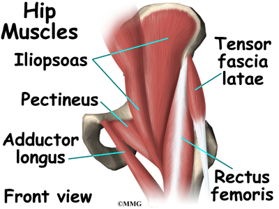
Anatomy Hip Muscles Diagram - Lower Back Muscle Anatomy And Low Back Pain : It is a prime mover of hip flexion and also acts to keep the upper body from falling.. Review muscle diagram using the 2 diagrams below: This mri hip joint axial cross sectional anatomy tool is absolutely free to use. Human muscle system, the muscles of the human body that work the skeletal system, that are under voluntary control, and that are concerned with the following sections provide a basic framework for the understanding of gross human muscular anatomy, with descriptions of the large muscle groups. Muscle and tendon anatomy of the hip (adductors, gluteal muscles (or buttocks), hamstring muscles, femoral muscle quadrices). The iliopsoas is a fused muscle composed of two muscles, the iliacus and the psoas major;
The ala provides an insertion point for the gluteal muscles laterally and the iliacus. Muscle and tendon anatomy of the hip (adductors, gluteal muscles (or buttocks), hamstring muscles, femoral muscle quadrices). Now that you watched the video, you. The muscles and fasciæ of the thigh. Click on the labels below to find out more about your muscles.
Hip Anatomy Eorthopod Com from eorthopod.com Now that you watched the video, you. Sartorius is a unique muscle because it is the only knee flexor that originates anteriorly. Knee assessment and hip mechanics online course: Microscopic anatomy of skeletal muscle. Muscle and tendon anatomy of the hip (adductors, gluteal muscles (or buttocks), hamstring muscles, femoral muscle quadrices). Click on the labels below to find out more about your muscles. The geometry of the hip allows wide range of motion in all planes, necessitating a large number of controlling muscles arising. They can be divided into three groups:
The accompanying muscle diagram reveals the positions of the muscles in this pose.
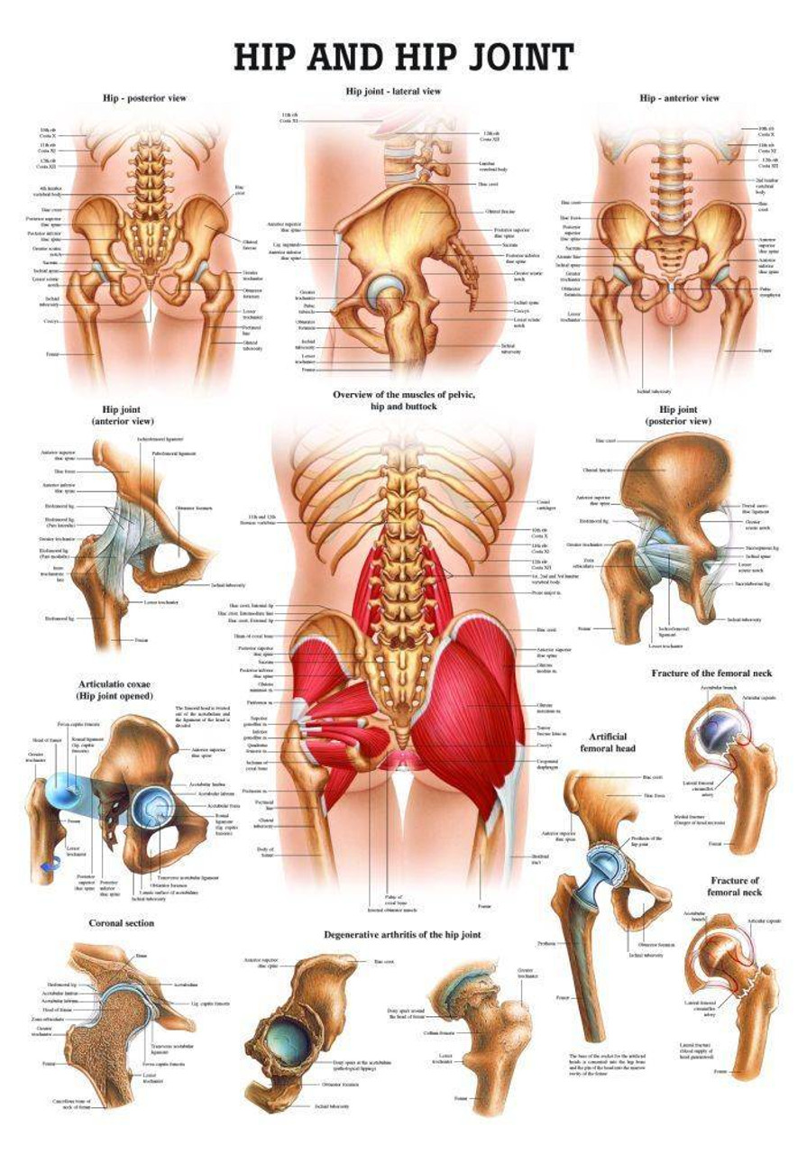
The hip joint is the articulation of the pelvis with the femur, which connects the axial skeleton with the lower extremity. Knowing the anatomy of your hip can help you understand the source of any hip pain. The geometry of the hip allows wide range of motion in all planes, necessitating a large number of controlling muscles arising. If you are starting to feel hip pain or stiffness, you'll want to know more about the bones and muscles that make up the hip's anatomy. Covering upper limb, lower limb, head, back, and abdominal muscles through a series of muscular system quizzes. Anatomical diagram showing a front view of muscles in the human body. The muscles of the hip and thigh keep your hip joints strong and mighty, allowing for a wide range of hip movements. David keil provides all you need to know about the. Most modern anatomists define 17 of these muscles. Download human muscle anatomy diagram vector art. The accompanying muscle diagram reveals the positions of the muscles in this pose. In the diagrams below, when you see muscle names that are the same color, it means they are an antagonistic pair and should not be both drawn first a few words about anatomy: Back muscles fitness gym muscle fitness fitness motivation health fitness health club.
Muscle tissue is also found inside of the heart, digestive organs, and blood vessels. Due to its muscular orientation, it causes flexion and lateral rotation at the hip. Diagram representing the anterior view of the muscle groups adductor brevis, adductor longus and adductor magnus. Knowing the anatomy of your hip can help you understand the source of any hip pain. Review muscle diagram using the 2 diagrams below:
Hip And Hip Joint Laminated Anatomy Chart from cdn11.bigcommerce.com Knowing the anatomy of your hip can help you understand the source of any hip pain. The hip joint is the articulation of the pelvis with the femur, which connects the axial skeleton with the lower extremity. Anatomy muscular system diagram human muscle stock muscular system back exercises anatomy and physiology. This mri hip joint axial cross sectional anatomy tool is absolutely free to use. Knee assessment and hip mechanics learn how hip. Microscopic anatomy of skeletal muscle. See more ideas about muscle diagram, medical anatomy, body anatomy. The different bursae of the hip region (trochanteric, ischial and.
*click them to make them larger & view details.
If you are starting to feel hip pain or stiffness, you'll want to know more about the bones and muscles that make up the hip's anatomy. Diagram representing the anterior view of the muscle groups adductor brevis, adductor longus and adductor magnus. The ilium is the largest part of the hip bone and makes up the superior part of the acetabulum. Inner hip muscles, outer hip muscles and muscles belonging to the adductor group 6. A collection of anatomy notes covering the key anatomy concepts that medical students need to learn. The geometry of the hip allows wide range of motion in all planes, necessitating a large number of controlling muscles arising. Skeletal muscle cells are multinucleate. Muscle tissue is also found inside of the heart, digestive organs, and blood vessels. They are a gland, so there is a hard mass in there, surrounded with soft fatty tissue. This mri hip joint axial cross sectional anatomy tool is absolutely free to use. Choose from over a million free vectors, clipart graphics, vector art images, design templates, and illustrations created by artists worldwide! Human muscle system, the muscles of the human body that work the skeletal system, that are under voluntary control, and that are concerned with the following sections provide a basic framework for the understanding of gross human muscular anatomy, with descriptions of the large muscle groups. The iliopsoas is a fused muscle composed of two muscles, the iliacus and the psoas major;
In the diagrams below, when you see muscle names that are the same color, it means they are an antagonistic pair and should not be both drawn first a few words about anatomy: The hip joint ( coxa in latin) is the articulation connecting the pelvis and the femur. Knowing the anatomy of your hip can help you understand the source of any hip pain. In this article, we will provide a basic overview of the muscles of the gluteal region while integrating clinical anatomical pathology to describe certain features. Muscle tissue is also found inside of the heart, digestive organs, and blood vessels.
Anatomy Hip Muscles Graph Diagram from graphdiagram.com The ala provides an insertion point for the gluteal muscles laterally and the iliacus. They can be divided into three groups: Click on the labels below to find out more about your muscles. In human anatomy, the muscles of the hip joint are those muscles that cause movement in the hip. David keil provides all you need to know about the. The hip abductor muscles play an important role in stabilizing the pelvis during gait, with its main function being performed by the gluteus medius. The different bursae of the hip region (trochanteric, ischial and. Thorough body movements (play gravity game using definitions).
Other sets by this creator.
This mri hip joint axial cross sectional anatomy tool is absolutely free to use. Review muscle diagram using the 2 diagrams below: Skeletal muscle cells are multinucleate. The different bursae of the hip region (trochanteric, ischial and. Knee assessment and hip mechanics online course: The muscles and fasciæ of the thigh. In human anatomy, the muscles of the hip joint are those muscles that cause movement in the hip. Download human muscle anatomy diagram vector art. The sacrum bone is almost always noticeable, no matter what the body type, because it the quadriceps muscles move the upper leg (femur) at the hip joint and the lower leg at the knee joint. The geometry of the hip allows wide range of motion in all planes, necessitating a large number of controlling muscles arising. If you are starting to feel hip pain or stiffness, you'll want to know more about the bones and muscles that make up the hip's anatomy. They are a gland, so there is a hard mass in there, surrounded with soft fatty tissue. And you'll be in a better position to help your doctor pinpoint the cause.
Knee assessment and hip mechanics online course: hip muscles diagram. Muscle and tendon anatomy of the hip (adductors, gluteal muscles (or buttocks), hamstring muscles, femoral muscle quadrices).